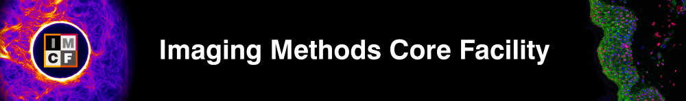
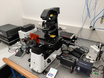
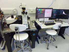
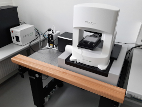
Carl Zeiss LSM 880 NLO
- Inverted confocal microscope
- Multiphoton excitation and non-descanned detection with FLIM option
- Internal spectral detection unit
Multi-functional fluorescence inverted confocal microscope with single- or multi-photon excitation, internal spectral detection and non-descanned detection with FLIM option.
Applications
- Fast and multi-color confocal imaging
- Various measurement options: z-stack, time series, tile scan, multi positions and regions
- Spectral detection, spectral unmixing and fingerprinting
- Deep imaging of thick biological samples with multiphoton excitation and non-descanned detection
- Label free imaging: Coherent anti-Stokes Raman Scattering (CARS) and higher harmonic generation (SHG and THG)
- FLIM with multiphoton excitation
- Long term live-cell imaging available
Specifications
Motorized inverted fluorescence microscope Carl Zeiss Axio Observer.Z1 with confocal module LSM 880 NLO equipped with the following:
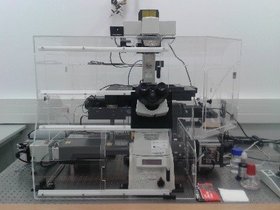
Nikon CSU-W1
- Spinning disk confocal microscope
- Dual camera, 7 laser wavelengths
- FRAP/Photoactivation module
- Heating/cooling stage top incubator
Versatile spinning disk confocal microscope equipped with seven excitation lasers, epifluorescence LED light source, and two back-illuminated sCMOS cameras. The microscope is equipped by stage-top heating/cooling incubator for live cell and tissue imaging and a FRAP/photoactivation module. It is based on an inverted widefield microscope Nikon Eclipse Ti2 equipped with motorized XY stage, Perfect Focus System, semi-motorized DIC, LED transmitted light source, multicolor LED for wide-field epifluorescence, piezo stage for fast z-stack acquisition, and automatic water dispenser for long term imaging.
Applications
- Fast multicolor confocal imaging
- Various measurement options: z-stack, time series, tile scan, multi positions
- Brightfield and DIC microscopy
- FRET imaging
Specifications
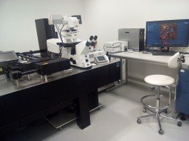
Abberior Instruments STED – INFINITY

- 3 dedicated 60x STED objectives (water, oil, and silicone)
- 5 excitation lines (405, 440, 485, 561, and 640 nm)
- Pulsed ultra-high-power STED laser (775 nm)
- 3 APD-based spectral detection channels
- 2 Spectral MATRIX detector (APD array) allowing differential detection on two channels
- TIMEBOW for measuring fluorescence lifetime with and without super resolution
- Adaptive optics for STED beams based on SLM technology
- Adaptive optics system (DFM deformable mirror) for all beams (excitation, emission, and depletion)
- Full auto-alignment
- Continuous hardware autofocus for STED & confocal
- Adaptive illumination package for highest resolution and live-cell super-resolution imaging at ultra-low light levels (RESCue,
- DyMIN, MINFIELD)
For more detailed microscope characteristics please see the section “Microscope”. Basic introduction to STED microscopy can be found here:
https://abberior-instruments.com/knowledge/microscopy-tutorials/sted/
Specifications
Software |
|
Epifluorescence | CoolLED pE-4000, 16 LEDs |
Epifluorescence cubes |
|
Excitation lasers |
|
STED Laser | 775 nm (40 MHz pulsed laser, for 2D and 3D STED) |
Donut formation | Programmable Spatial Light Modulator – 775 nm |
Scanner | Abberior QUAD scanner |
Objectives |
|
Rainbow Spectral Detection Kit |
|
Pinholes | 16 pinholes from 25 μm to 2 mm |
Detectors |
|
FLIM |
|
RAYSHAPE Adaptive Optics system |
|
FLEXPOSURE Adaptive Illumination |
|
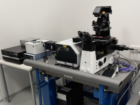
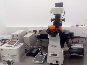
Abberior Instruments STED
- Inverted confocal microscope
- STED super-resolution microscopy
Fluorescence inverted confocal microscope and STED super-resolution nanoscope with high detection efficiency of far-red photons. The system is equipped with four excitation lasers, depletion laser 775 nm and four single photon counting detectors. For more detailed microscope characteristics please see the section “Microscope”. Basic introduction to STED microscopy can be found here:
https://abberior-instruments.com/knowledge/microscopy-tutorials/sted/
Applications
- Two-color 2D and 3D super-resolution images obtained by STED technique with a pulsed depletion laser 775 nm and pulsed excitation 561 nm and 640 nm
- STED RESCUE and DYMIN mode available (STED imaging mode that significantly reduces the light dose sent onto the sample without compromising the resolution)
- 2D and 3D STED images available also with water immersion objective
- Multicolor confocal scanning system with variable pinhole size
- Possible FLIM or FCS acquisition
- Live cell imaging
- NIR confocal imaging (775 nm excitation)
Specifications
Inverted confocal microscope Nikon Eclipse Ti-E equipped with a piezo Z-stage, motorized XY stage, Perfect Focus System, transmitted light lamp (100 W) and following units:
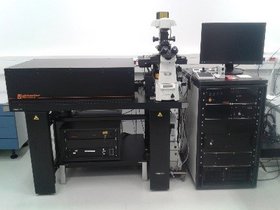
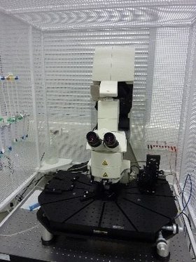
Carl Zeiss Lattice Lightsheet 7

- Short term, long term live cell imaging at subcellular resolution
- Low phototoxicity and photobleching
- Dual camera setup for colocalization studies
- High speed volumetric imaging, penetration depth up to 200 µm
- Tissue culture cells, cells in hydrogel up to spheroids and organoids
Lightsheet microscope with gentle light sheet imaging at high resolution and volumetric speed with standard sample carriers. Complex alignment processes are performed automatically on workstation with high-speed data transport connection.
Applications
Volumetric live cell imaging of:
- adherent or suspension cells
- 3D cell cultures: cysts, cells in hydrogel, organoids, spheroids with diameter up to 200µm
- Small evolving organisms: zebrafish embryo, C.elegans embryo, Drosophila embryo
Specifications
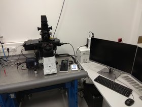
Nikon iLas 2 ring-TIRF/FRAP
- Inverted widefield microscope
- ring-TIRF illumination
- Image splitter (optional)
- Two cameras (sCMOS, EM-CCD)
Inverted fluorescence widefield microscope based on Nikon ECLIPSE Ti2 microscope body and iLas 2 (Gataca Systems) illumination device allowing ring-TIRF microscopy and FRAP.
Applications
- Fast multicolor widefield imaging with TIRF or HILO excitation option
- Bright-field microscopy
- Epi-fluorescence
- Interference reflection microscopy
- FRAP (fluorescence recovery after photobleaching) measurements
- Option of simultaneous dual wavelength imaging by single camera
- Long term live-cell imaging available
Specifications
Inverted widefield microscope body Nikon Eclipse Ti2, motorized XY stage, Perfect Focus System, automatized H-TIRF module, insertable gradation neutral density filter, transmitted light lamp (100 W) and following units:
Nikon N-STORM
- Inverted widefield microscope
- TIRF illumination
- Image splitter
Multifunctional fluorescence inverted widefield microscope enabling live-cell imaging, TIRF or HILO illumination, and single molecule localization microscopy (SMLM) superresolution technique.
Applications
- Single Molecule Localization Microscopy methods (dSTORM, PALM, DNA-PAINT)
- Two-color dSTORM images acquired by spectral demixing
- Fast and sensitive multicolor widefield imaging with TIRF or HILO excitation option
- Option of simultaneous dual wavelength imaging by single camera
- Long term live-cell imaging available
- Brightfield microscopy
Specifications
Inverted widefield microscope Nikon Eclipse Ti-E equipped with a piezo Z-stage, motorized XY stage, Perfect Focus System, automatized H-TIRF module, insertable quarter-wave plate, insertable gradation neutral density filter, transmitted light lamp (100 W) and following units:
Nikon Ti-E H-TIRF
- Inverted widefield microscope
- TIRF illumination
- Image splitter
Versatile fluorescence inverted widefield microscope equipped with four lasers, epifluorescence lamp and EM-CCD and CMOS cameras. The microscope is designed for short and long live-cell imaging and experiments under standard or TIRF illumination. Link to manufacturer website:
https://www.nikoninstruments.com/cz_CZ/Vyrobky/Inverzni-mikroskopy/Eclipse-Ti-E
Applications
- Fast and sensitive multicolor widefield imaging with TIRF, HILO or EPI excitation option
- Option of simultaneous dual wavelength imaging by single camera
- Short or long term live-cell imaging available
- Various measurement options: z-stack, time series, tile scan, multi positions
- Brightfield and DIC microscopy
Specifications
Inverted widefield microscope Nikon Eclipse Ti-E equipped with motorized XY stage, Perfect Focus System, H-TIRF module, module for environmental control Okolab (temperature controlled range room temperature up to 40°C; CO2 0-10%; humidity up to 95 %), insertable quarter-wave plate, insertable gradation neutral density filter, transmitted light lamp (100 W) and following units:
Nikon Ti-E H-TIRF 2
- Inverted widefield microscope
- TIRF illumination
Versatile fluorescence inverted widefield microscope equipped with three lasers, epifluorescence lamp, and back-illuminated sCMOS camera. The microscope is designed for short- and long-term imaging experiments under standard or TIRF illumination. Links to manufacturer website:
https://www.microscope.healthcare.nikon.com/products/inverted-microscopes/eclipse-ti2-series
Applications
- Fast and sensitive multicolor widefield imaging with TIRF, HILO or EPI excitation option
- Various measurement options: z-stack, time series, tile scan, multi positions
- Brightfield and DIC microscopy
Specifications
Inverted widefield microscope Nikon Eclipse Ti2 equipped with motorized XY stage, Perfect Focus System, H-TIRF module, insertable quarter-wave plate, insertable gradation neutral density filter, transmitted light lamp (Nikon TI2-D-LHLED) and following units:
Leica TCS SP8 WLL SMD-FLIM
- Inverted confocal microscope
- WLL and internal spectral detection unit
- FLIM and FCS
Versatile inverted fluorescence confocal microscope equipped with 405 nm, 445 nm and white light laser, spectrally tunable detection and FLIM hardware. Presence of camera and epifluorescence lamp allows also widefield imaging. For more detailed microscope characteristics please see the section “Specifications”.
Applications
- Multi-color confocal imaging
- Multi-color widefield imaging
- Various measurement options: z-stack, time series, tile scan, multi positions
- Spectral imaging
- Measurement of emission and excitation spectra
- Lifetime imaging, FLIM-FRET
- Measurement of e.g. membrane dynamics by FCS and FRAP
- Live-cell imaging available: stage-top incubator with controlled humidity and atmosphere (CO2+N2)
- Brightfield, DIC, polarized light microscopy
Specifications
An inverted confocal microscope Dmi8 with a laser scanning confocal head Leica TCS SP8, a motorized microscope stage with a Super Z-galvo scanning insert for fast and precise Z movement, HW autofocus and Best focus control, transmitted light LED U-12V, square pinhole of tunable size and following units:
Leica TCS SP8 DM6 CFS
- Upright confocal microscope
- Internal spectral detection unit
An upright fluorescence confocal microscope equipped with four excitation lasers, dipping objectives with long working distance, oil immersion objective and 1x HyD detector and 2x PMTs. Presence of camera, epifluorescence lamp and removable infrared filter allows also widefield imaging in infrared area. Accessory for electrophysiological experiments is also available. For more detailed microscope characteristics please see the section “Specifications”.
Applications
- Fast and multi-color confocal imaging
- Fast and multi-color widefield imaging
- Various measurement options: z-stack, time series
- Spectral imaging, measurement of emission spectrum
- Option of infrared detection in widefield imaging
- Brightfield and polarized light microscopy
Specifications
An upright confocal microscope DM6 CFS with a laser scanning confocal head Leica TCS SP8, a manual microscope stage, Best focus control, transmitted light LED U-12V, removable infrared filter, square pinhole of tunable size and following units:
Leica DMi8 WF
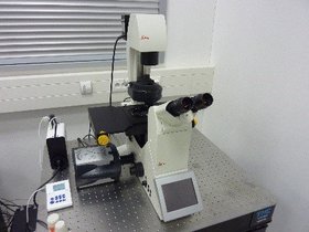
- Inverted widefield microscope
Inverted widefield fluorescence microscope equipped with epifluorescence lamp and EM-CCD camera.
Applications
- Fast and multi-color widefield imaging
- Various measurement options: z-stack, time series, tile scan, multi positions
- Brightfield and DIC microscopy
Specifications
An inverted widefield microscope Dmi8 with motorized microscope stage, transmitted light LED U-12V, and following units:
Carl Zeiss Axiozoom.V16
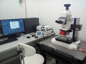
- Upright macroscope
Fluorescence zoom upright microscope which combines the benefits of stereomicroscopes such as zoom optics and long working distances with the higher resolutions of traditional light microscopes. The macroscope in IMCF at BIOCEV is equipped with a 2.3x magnification objective combined with numerical aperture 0.57 what offers superior brightness in large objects fields. In comparable image fields, the Axio Zoom.V16 offers a 2.5 times higher resolution and 10 times brighter fluorescence than generally used stereomicroscopes. See the product web page.
Applications
- visualization of large samples, e.g. whole model organism, in fluorescence contrast or transmitted light with the possibility to zoom in to the smallest details of the sample. Long working distance of the available objectives allows comfortable manipulation with specimens.
- basic documentation of histological samples.
Specifications
Axiozoom.V16 is a fluorescence zoom upright microscope equipped with a motorized stage with travel range 150 x 100 mm and the following:
Akoya PhenoCycler-Fusion 2.0

PhenoCycler-Fusion 2.0 enables to detect protein expression on tissues in real time. The system introduces up to 64 cycles automation with rapid fluidics establishing itself as the fastest spatial biology solution enabling to detect up to 100+ biomarkers (3 markers per run plus DAPI as reference). It is a part of an integrated end-to-end workflow containing pre-optimized antibodies and reagents, fast fluidics and imaging, as well as onboard data processing generating high-resolution QPTIFF files. The system features imaging of 1 million cells in 10 minutes by the detection of biomarkers while preserving tissue integrity using barcoding technology. The system allows to stain the tissue also with non-inventoried antibodies conjugated with barcodes via kit. Panel could be built up from an extensive database of antibodies, in-house conjugated antibodies or Akoya offers ready to-use PhenoCode™ Discovery Panels
Features
- For FFPE and FF tissue sections, tissue microchips, tissue sections, organoids
- Up to 100+ biomarkers on one section by unique barcoding technology
- Up to 300+ catalog antibodies or in-house conjugated antibodies
- Cell phenotyping
- Cellular neighborhood analysis
- Slide scanning without multiplexing, brightfield, and fluorescence
- 25 min of acquisition for 4 colors, at 15 mm x 15 mm, at 20x
- QPTIFF file format compatible with Enable Medicine, Visiopharm, QuPath etc.
Specifications
Nanolive CX-A
- 3D holotomography & fluorescence
- Stage-top incubator for long-term automatic live cell imaging
Automated holotomographic and fluorescence microscope for long-term live-cell imaging. Label-free 3D imaging of cells in real time is ensured due to the low power laser and top stage. In addition, presence of epifluorescence lamp allows correlative holographic and fluorescence imaging. Link to manufacturer website:
https://www.nanolive.ch/products/3d-microscopes/cx-a/
Applications
- Gentle long-term live-cell label-free 3D imaging
- Correlative holographic (3D) and fluorescence (2D) imaging
- Imaging of cell populations, single cells or organelles
- Measurement of refractive indices inside the cell
Specifications