Electron microscopes
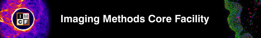
FEI Helios NanoLab 660 G3 UC
- FIB-SEM DualBeam microscope
- High resolution scanning electron microscopy with focus ion beam
Scanning electron microscope with focused ion beam milling equipped for cryo-imaging and correlative light and electron microscopy (CLEM)
Applications
- 3D scanning electron microscopy (FIB-SEM)
- high resolution scanning electron microscopy (embedded, dried or frozen samples)
- elemental analysis (EDS)
Specifications
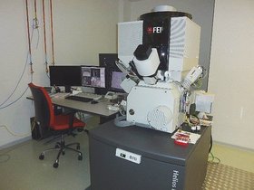
JEOL JEM 2100-Plus 200kV
- Transmission Electron Microscope equipped with cryo-pole piece and on axis TVIPS TemCam–XF416 4K CMOS camera with frame rate up to 24 f/s and high sensitivity
Transmission Electron Microscope equipped with a cryo-pole piece, fast and sensitive TVIPS XF-416 4kx4k camera and side-entry cryo holder Gatan 626.
Applications
- Transmission electron microscopy (TEM) at room temperature
- Advanced sample mapping
- Electron tomography (ET)
- Diffraction at cryo and room temperature
- Cryo-electron microscopy
- SerialEM software for automated image acquisition, montage mapping, tomography in ambient and cryo conditions
Specifications
Sample preparation equipment
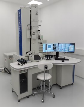
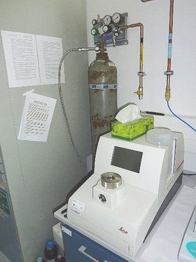
Automatic Plunge Freezer EM GP2
- The EM GP2 plunge freezes fluid or extremely thin samples spread on an electron microscopy grid into liquid ethane after removing excess fluid by automatic blotting.
- Prior to freezing the sample is maintained in a temperature and humidity controlled environmental chamber, adjustable between +4°C and +60°C and room humidity to 99 %
Specifications
Leica EM GP2 on Leica Microsystems webpage (https://www.leica-microsystems.com/products/sample-preparation-for-electron-microscopy/p/leica-em-gp2/)
Leica EM GP2 Flyer (https://www.leica-microsystems.com/products/sample-preparation-for-electron-microscopy/p/leica-em-gp2/)
Leica EM ICE – high pressure freezing machine
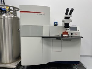
- cryofixation of samples for EM
High pressure freezing system for cryo-fixation (vitrification) of samples for EM. There are several advantages over chemical fixation. Sample is freeze in a range of milliseconds, which immobilize all macromolecular components simultaneously. This technique also improves preservation of biological samples.
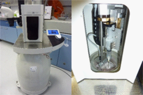
Leica EM ACE600 – coating system

- precise coating of samples foe subsequent examination with an electron microscope
- glow discharge
- freeze fracture cryo set
Application
- Metal coating of sample to cerate a conductive layer which inhibits charging, reduces thermal demage and improves the secondary electron signal in the SEM
- Making the carbon layers to be transparent to the electron beam but conductive (needed for X-ray microanalysis), to support films on grids
- Glow discharge to render grids surface hydrophilic
- Freeze-Fracture of frozen samples
Main components
The Leica EM ACE600 includes the following main functional units:
- High vacuum chamber
- Rotation sample stage, 24 positions for 12,7 mm SEM stubs
- Quartz thickness measurement
- Carbon thread source or sputter source (platinum and gold targets)
- Freeze-Fracture cryo set includes vacuum cyro transfer Leica EM VCT100 and cryo stage
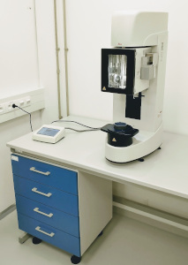
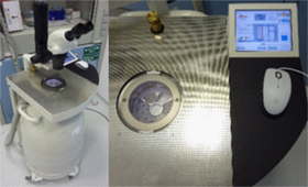
Leica EM CPD300 – critical point dryer
- drying of samples with CO2 in critical point for SEM
Automated Critical Point Dryer
Application
- The drying of biological specimens such as pollen, tissue, plants, insects for SEM analysis
- The drying of industrial samples , for example MEMS (Micro Electro Mechanical Systems)
Method
Crytical point drying is a method which use of CO2 as transitional fluid because its critical point is at 31 °C and 73,8 bar. Technically these temperature and pressure requirements of the CO2 can be implemented relatively easily then for water (critical point is 374 °C and 228,5 bar). The water in the cell is replaced by an acetone which is very soluble with liquid CO2. This procedure is follow by substitution with liquid CO2 through serial dilution steps. The increasing the temperature and pressure will transfer the CO2 through its critical point into a subcritical state. By controlled depressurization and constant temperature is subcritical CO2 convert into its gaseous phase without crossing the phase boundary between liquid and gas. This technique of preparing sample for scanning electron microscope is more gentle and better preserve structure than air drying sample.
Leica EM AFS2 & FSP (freeze substitution processor)
- An automatic reagent handling system for AFS2
Automatic freeze substitution (AFS) connects cryo-fixation (e. g. high pressure freezing) and resin embedding. It is a process of dehydration in low temperatures without crystal formation. In the beginning of freeze substitution, amorphic water contained in sample is dissolved by an organic solvent (e.g. acetone, ethanol), which also includes chemical fixatives. After dehydration temperature rises to point where is resin embedding possible. Freeze substitution processor (FSP) is an automatic reagent handling system combined with the Leica EM AFS2, dispenses reagents for freeze substitution. It also provides UV lamp for photopolymerization of acrylic resins.
Application
Cryo-substitution and resin embedding
Types of sample |
|
Resins |
|
Leica EM AFS2 (automatic freeze substitution)
- Cryo-substitution for samples frozen in HPM100
Automatic freeze substitution (AFS) connects cryo-fixation (e. g. high pressure freezing) and resin embedding. It is a process of dehydration in low temperatures without crystal formation. In the beginning of freeze substitution, amorphic water contained in sample is dissolved by an organic solvent (e.g. acetone, ethanol), which also includes chemical fixatives. After dehydration temperature rises to point where is resin embedding possible. Freeze substitution processor (FSP) is an automatic reagent handling system combined with the Leica EM AFS2, dispenses reagents for freeze substitution. It also provides UV lamp for photopolymerization of acrylic resins.
Application
Cryo-substitution and resin embedding
Types of sample |
|
Resins |
|
Pelco BioWavePro+ – microwave processor
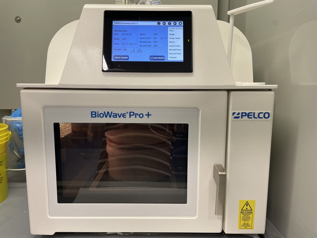
- sample preparation for EM
Microwave Processor containing Pelco ColdSpot®Pro cooling system
Application
Sample preparations for EM (fixation, contrastation, dehydratation, embedding, polymerisation)
Types of sample |
|
Adjustable features |
|
On/Off systems |
|
Other features |
|
Ultramicrotome Leica EM UC7
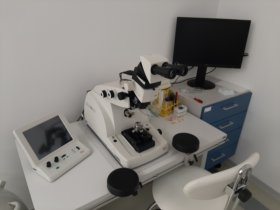
- EM UC7 Basic Instrument with eucentric 180° swivelling microscope carrier
- Built in anti-vibration system
- Cutting window adjustment from 0.2-15 mm
- Cutting window adjustment from 0.2-15 mm
- Knife block 360° rotatable, dual sided self locking precision drive with ±30° graduation, clearance angle drive with 1° scale from -2° to over 15°
- Knife holder for 6-12mm glass and diamond knives
- 360° rotation of specimen with self locking drive
Specification
- Eucentric movement of the stereo microscope carrier (+ 5° / – 8°) with patented designated positions for specimen approach, for glass and diamond knives
- Multi-LED-illumination for standard illumination, back light illumination, specimen
transillumination and spot light illumination - Dual automatic advance system with stepping motor for 200μm specimen advance, stepping motor for 10mm knife N-S movement
- Stepping motor for 25mm E-W movement
- Reflexomat II -peristaltic water pump; water spout in magnetic holder
- Syringe, special tubing for Reflexomat II
- Segment arc with specimen transillumination, self locking precision drives with ±22° eucentric movement, transferable stem for longitudinal and cross sectioning
Stereomicroscope Leica Investa 3 (Integrated camera)

- Designed to record and facilitate sample dissection and micromanipulations, preparation and quality control of sample carriers a tools
- Optimized for stereomicroscopic inspection of samples, acquisition of documentation images/movies, demonstrations and user training
Specification
- Integrated camera and output to an external monitor are suited for digital image sharing directly on different devices incl. external monitor, mobile device, or computer.
- Enables 3D view of the sample with an optimal 3D perception thanks to FusionOptics which combines high resolution and a large depth of field, large 9:1 zoom range, magnification of up to 55x and apochromatically corrected optics.
- Convenient to handle samples with the microscope’s extra large working distance up to 122 mm.
- Enersight software interface can be used in multiple operation modes, such as on-screen display, mobile devices, or computer, allowing for inspection, comparative analysis and length/angle measurements.
- Integrated camera can visualize sample movements on the monitor clearly and smoothly thanks to the high frame rate (up to 60 fps) with the fast auto exposure in the live image display.
- Leica Investa 3 Flyer (link)