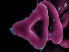
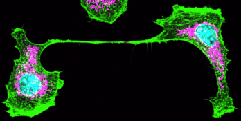
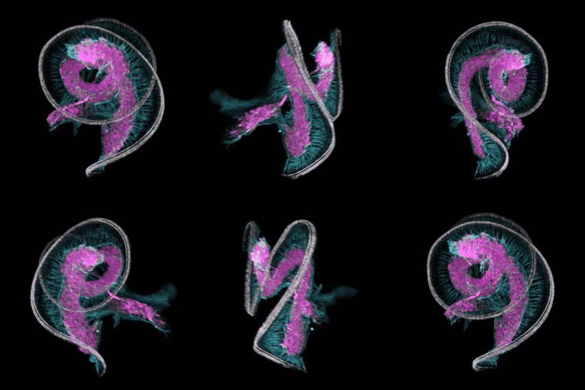
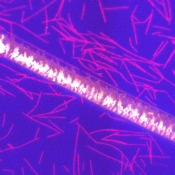
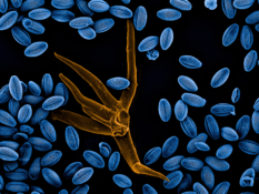
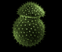
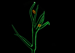
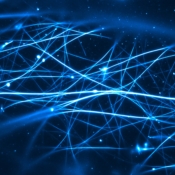
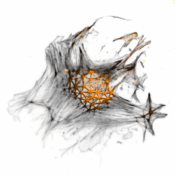
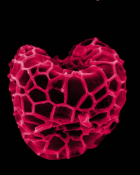
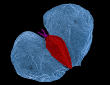
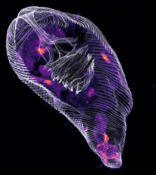
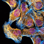
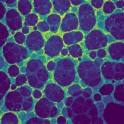
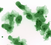
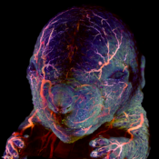
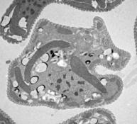
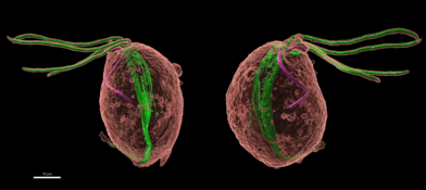
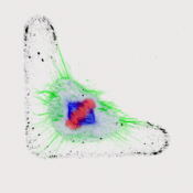
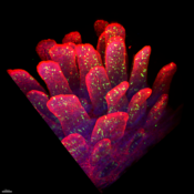
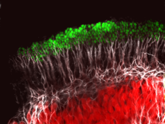
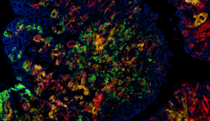
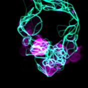
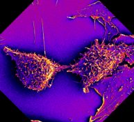
Monthly Winners Click on image for higher resolution! Alexandre Beber: Labelled actin filaments (in yellow) and labelled Anillin + RhoA (in blue) have been deposited on the surface because the buffer dried out. This was not intentional but looks pretty nonetheless. Ashley Hannah George: Vitrified sample of DLPC liposomes visualised by cryo-EM technique. Michaela Ždímalová: Tongues of fire; A network of actin filaments (orange) and microtubules (red) contracted by an over-concentrated myosin II (unlabelled). Dominik Arbon: Acanthamoeba phagocytosis, Acanthamoeba castellanii with fluorescently tagged vacuoles (green) phagocyting Escherichia coli (red). The image was used as cover art for the International Journal of Parasitology, July 2022. Adéla Karhanová: Leave from a different world; Drying channel with fluorescent tau protein on microtubules. Jitka Kučerová: Fantastic trichomonads and where to find them; Trichomonas vaginalis cell stained with antibody against tubulin. Ravi Kumar: Live imaging of anaerobic Trichomonas vaginalis-Iron sulfur cluster U (IscU) protein used as Hydrogenosomal marker by tagging with Photoactivable Yellow protein (Pyp). Miles Raishbrook: Visualising the site of phototransduction. TEM image of a mouse’s rod photoreceptor outer segments (pseudocoloured to delight your cones). Sebastian Cristian Treitli: Polymastix sp. with various types of surface bacterial symbiont. A cell of Polymastix with its nucleus shown in blue and two types of surface bacterial symbionts highlighted in purple and green. Eliška Miková: Fatal attraction: Axonal growth cone seeking its cue Martina Vinopalová: In the expanded world of Giardia Localization of the GiMlf (green) protein in a stack of images of an expended cell of Giardia intestinalis, costained with a NHS-ester dye.